Heels of Progress
Posted May 2012 in History of Photography, Scientific Photography
If you ever wanted to learn about the importance assigned or excitement surrounding the discovery of the X-Ray, look no further than any photographic journal published the world over between 1896-1897. Chronicled in breathless detail within their many pages, this new and miraculous revelation was aided by photography’s very ability to record the see-through results of these “mysterious rays” on a myriad of materials.
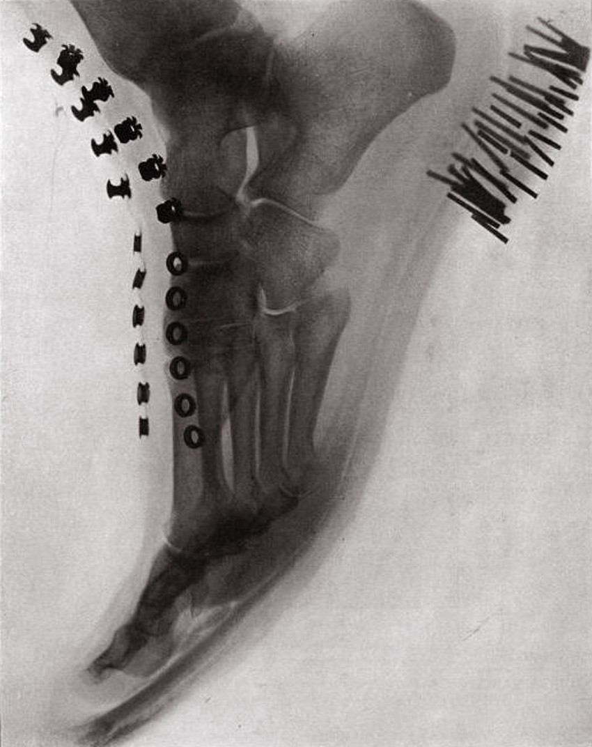 "Foot in a Shoe": full-page halftone plate identified as figure 6 accompanying article "Radiography and its Application" published in "The Photographic Times": July: 1896. Believed to be photographed by author Arthur Willis Goodspeed with the assistance of G. C. McKee.
And so this new victory was shouted far and wide: the symbolic Iron Heel of Progress, represented by the dual disciplines of scientific investigation and photography coming together, marched forward. In my own convoluted way of thinking, the splendid specimen of shoe including said iron-studded heel protecting a foot within makes perfect sense, literally and perhaps symbolically making a full-page debut along with other objects in the July, 1896 issue of The Photographic Times.
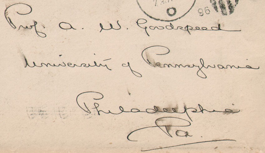 University of Pennsylvania physics professor and Radiology pioneer Arthur Willis Goodspeed was the addressee of this personal letter sent by Photographic Times editor Walter E. Woodbury in 1896 seeking the procurement of X-Ray photographs to accompany Goodspeed's published July issue article: "Radiography and its Application". Detail from original envelope with dimensions of 9.3 x 16.5 cm
The reason for all this excitement was the official announcement late the year before: German physicist Wilhelm Röntgen (1845-1923) had “produced and detected electromagnetic radiation in a wavelength range today known as X-rays or Röntgen rays”. (1.) For his efforts, Röntgen in 1901 was awarded the first Nobel Prize in Physics.
I will be the first to admit scientific photography is not a collecting focus for the PhotoSeed archive, however, the possession of a postal cover and several pages from a hand-written letter by Photographic Times editor Walter E. Woodbury (1865-1905) posted to this site was reason enough to visually explore X-Ray photography in this space as the profound discovery it remains even today. On May 22nd, while working in advance of the July issue, Woodbury penned a short missive to America’s equivalent of Röntgen: University of Pennsylvania physics professor Arthur Willis Goodspeed. (1860-1943) Radiography and its Application was the name of the article he had already written, dated April 30th and eventually published. But at the time, working more than a month in advance, editor Woodbury was willing to hold up publication of his journal until he could secure the necessary photographs showing the dry-plate, x-ray-effected negatives he knew would cause a stir, and thus providing proof for and generating interest in Goodspeed’s article.
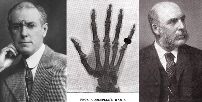 Left: Arthur Willis Goodspeed (1860-1943) circa 1903-04 when he was 4th President of the American Roentgen Ray Society. Middle: An X-Ray photographic negative from 1896 showing Goodspeed's hand taken by Philadelphia photographer John Carbutt. Right: Englishman John Carbutt, (1832-1905) inventor of specialized glass dry plates sensitive to the newly identified x-rays that were provided to Goodspeed for research purposes.
Goodspeed was no stranger to photographic experimentation. In the mid 1880’s he had witnessed and assisted the English photographer Eadweard Muybridge (1830-1904) while he conducted the now famous Animal Locomotion studies under the support of the University of Pennsylvania and more unbelievably, had made by accident along with British photographer William Jennings, the first known X-Ray photograph in the physical lecture room at the school on February 22, 1890 . A centennial remembrance written by TL Walden Jr. for the journal Radiology in 1991 partly states:
On that evening, Goodspeed and Jennings had been making brush electrographs of coins and brass weights. After they finished their experiments, Jennings stacked all of the photographic plates; two coins—either left from the experiments or Jennings’ trolley fare—were placed on top of the plates. Goodspeed then demonstrated to Jennings the university’s collection of Crookes tubes, with the idea of photographing the glow from the tube. While the two men were talking, however, the Crookes tube was emitting x radiation that affected the nearby plates. After the plates were developed, Jennings noted that one had the shadow(s) of a disk(s) on it; neither man could explain the image. (2.)
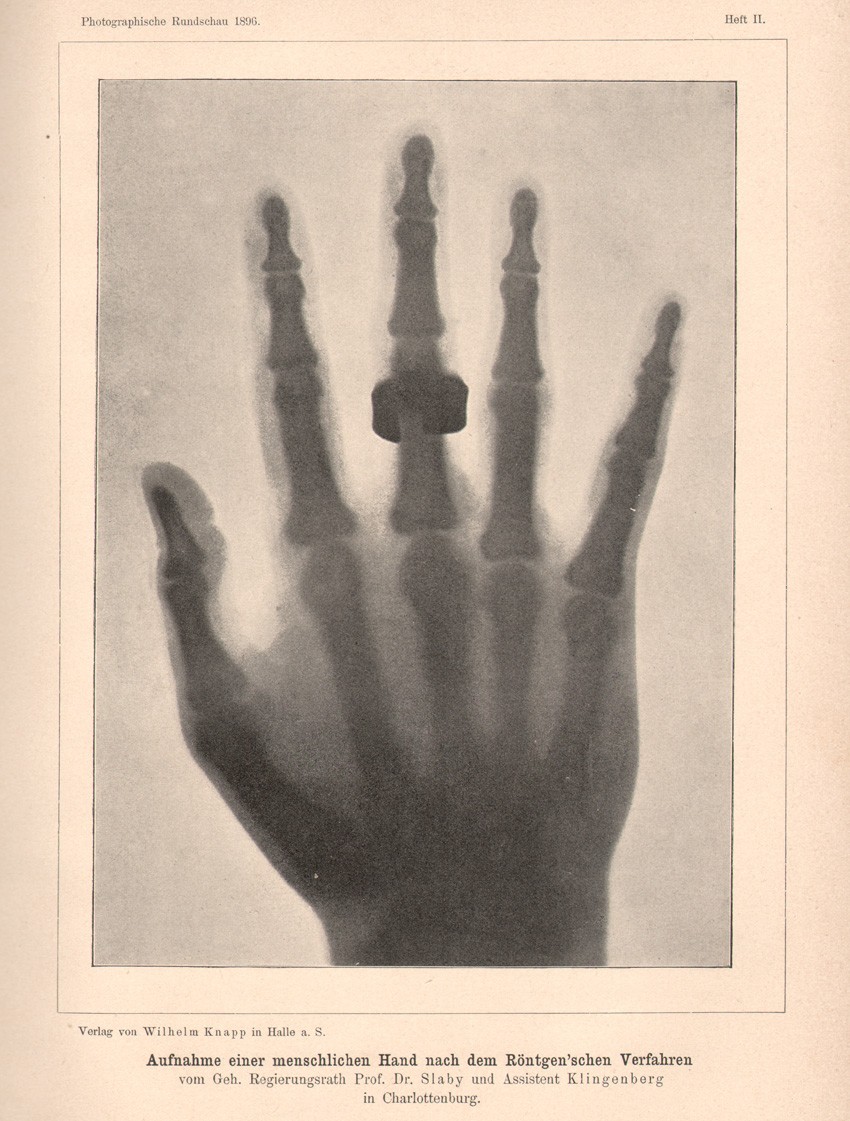 One of the most popular subjects it seems for early depictions of X-Ray negative photographs was the human hand. This full-page halftone presentation of a full hand with ring was included in the February, 1896 issue of the German photographic journal Photographische Rundschau. Original caption: Aufnahme einer menschlichen Hand nach dem Röntgen'schen Verfahren vom Geh. Regierungsrath Prof. Dr. Slaby und Assistent Klingenberg in Charlottenburg.
The photographic holdup for Woodbury was worth it. Englishman John Carbutt, (1832-1905) who had first made a name for himself in America by taking stereoscopic landscape photographs as well as running a Chicago portrait studio in the 1860’s, had become an important collaborator in the late 1890’s with Goodspeed in Philadelphia. Carbutt’s invention of specialized glass dry plates sensitive to the newly identified x-rays were provided to Goodspeed for research purposes; the same year his article appeared in The Photographic Times. Carbutt’s role as well as the importance of these plates was acknowledged in it:
With a view to developing the sensitive plate to produce the best results possible, Mr. John Carbutt has given untiring attention and made many experiments. The Carbutt plates have most of them been tested by the writer in comparison with other makes, and those now in use give by far the best results of any yet tried. The negatives from which the illustrations accompanying this article have been reproduced are samples of the plates referred to. (3.)
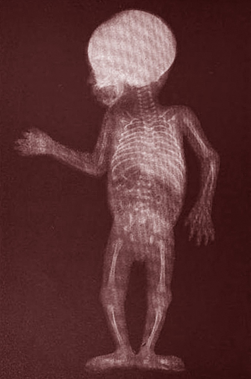 Unknown health hazards did not seem to present issues with photographers keen to exploit the miracle that was X-Ray photography when first discovered in late 1895. Although it is not known what the exposure time for this 3 day old child was when Philadelphia photographer John Carbutt recorded it in 1896, exposures of over 1 hour in length are commonly mentioned. This photograph appeared as a full-page halftone in the December, 1896 issue of "The American Amateur Photographer".
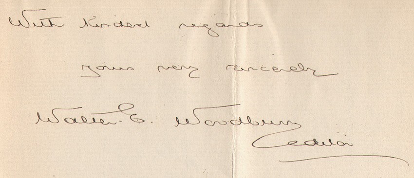 "Photographic Times" editor Walter Edward Woodbury (1865-1905) was the son of Walter B. Woodbury, who invented the Woodburytype. Woodbury edited the journal from 1895-1899. He died from yellow fever while later editing the English section of the "Panama Star and Herald and Inter-Ocean Critic" newspaper in Panama.
In closing, and with a nod to collectors like myself seeking out the ultimate published examples of Röntgen, or X-Ray scientific photographs, I suggest a further investigation of the 15 oversized, hand-pulled photogravure plates published in 1896 under the direction of Austrian photo-chemists Josef Maria Eder and Eduard Valenta. Containing magnificent studies of human bones, various small animals as well as man-made objects including a set of lockets, this portfolio, titled Versuche über Photographie mittelst der Röntgen’schen Strahlen, features as its’ final plate the now iconic coiled snake titled Aesculap-Schlange. First taken by Eder and Valenta and presented to members of the Viennese Photographic Society in January of 1896, (4.) these photographs have long ago entered the canon of modern photographic art, a scant two months after Röntgen’s initial discovery shook the world.
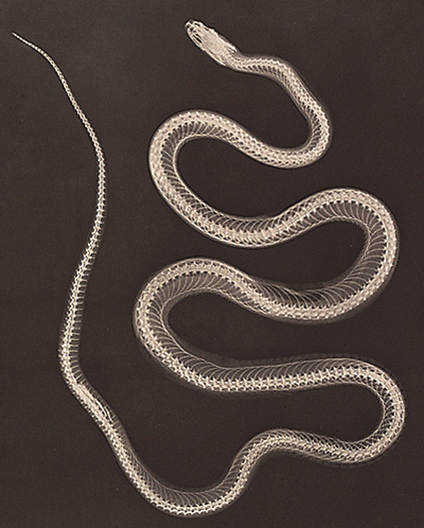 Detail: Aesculap-Schlange (Facsimile des Negativs). pl. XV: from portfolio: "Versuche über Photographie mittelst der Röntgen'schen Strahlen" published in 1896 as a large plate photogravure. Symbolic of healing and native to Europe, the Aesculapian snake is associated with the Greek god Asclepius and Roman god Aesculapius. The symbol of modern human medicine is often represented by this snake intertwined around a rod.
1. Wilhelm Röntgen: from: Wikipedia: accessed: 2012
2. excerpt: The first radiation accident in America: a centennial account of the x-ray photograph made in 1890: TL Walden Jr.:in: Radiology: December, 1991: pp. 635-639
3. excerpt: Radiography and its Application: A.W. Goodspeed: in: The Photographic Times: New York: July, 1896: pp. 308-309
4. from: Beauty of Another Order-Photography in Science: Ann Thomas: Yale University Press: New Haven and London, in association with the National Gallery of Canada, Ottawa; 1997
This entry was posted on Thursday, May 3rd, 2012 at 12:44am and is filed under History of Photography, Scientific Photography. You can follow any responses to this entry through the RSS 2.0 feed.